Course: High school biology > Unit 2. Lesson 2: Basic cell structures. Introduction to the cell. Introduction to cilia, flagella and pseudopodia. Basic cell structures review. Identifying cell structures. Basic cell structures. Science >. High school biology >.
Stem cell classification and its application | MedChemExpress
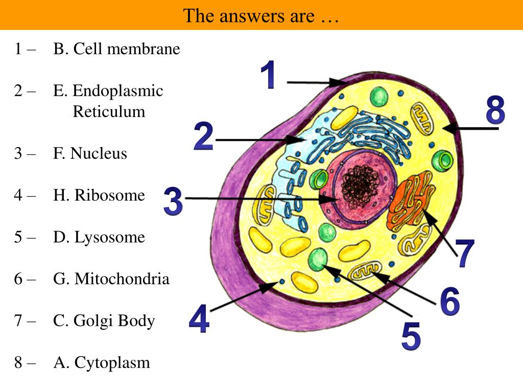
Students practice labeling organelles on a simple model (2D) and a more complex model. The idea is for students to gain an appreciation for how cell diagrams are created. They don’t all look alike, and are often artistically created. Cell organelles tend to follow basic design rules, like the mitochondria will generally look like a peanut

Source Image: slideserve.com
Download Image
Draw two adjacent pepper epidermal cells. Label the cell wall, middle lamella, plasmodesmata, and chromoplasts. You are encouraged to identify and label other cell components, such as the nucleus and nucleolus, if they are visible. A potato is a modified part of the plant called a tuber. Much like an onion, a tuber is a part of the plant–this

Source Image: brainly.com
Download Image
PARTS OF THE CELL. Label the numbered parts. Then complete the table by writing down the differences 2. – Brainly.ph 1. Plasma membrane: a selective barrier which encloses a cell (plant and bacteria cells also contain a cell wall ). 2. Cytosol: located inside the plasma membrane, this is a jelly-like fluid that supports organelles and other cellular components. 3. Cytoplasm: the cytosol and all the organelles other than the nucleus. 4.

Source Image: pubs.acs.org
Download Image
Label The Two Cell Parts On The Diagram Below.
1. Plasma membrane: a selective barrier which encloses a cell (plant and bacteria cells also contain a cell wall ). 2. Cytosol: located inside the plasma membrane, this is a jelly-like fluid that supports organelles and other cellular components. 3. Cytoplasm: the cytosol and all the organelles other than the nucleus. 4. 1. Write the name of the cell part in the box next to its description/function. Site of protein synthesis; attached to the outside surface of the endoplasmic reticulum; produces proteins for use outside the cell or for use in the cell membrane. 2. Indicate if each of the following is true of chromosomes or chromatin.
Monitoring Protein Import into the Endoplasmic Reticulum in Living Cells with Proximity Labeling | ACS Chemical Biology
Jun 6, 2023Animal cell size and shape. Animal cells come in all kinds of shapes and sizes, with their size ranging from a few millimeters to micrometers. The largest animal cell is the ostrich egg which has a 5-inch diameter, weighing about 1.2-1.4 kg and the smallest animal cells are neurons of about 100 microns in diameter. Year 9 biology cells Flashcards | Quizlet

Source Image: quizlet.com
Download Image
In the diagram below, Cell I and II represent typical cells. In both cells, organelle 5 is the site of – brainly.com Jun 6, 2023Animal cell size and shape. Animal cells come in all kinds of shapes and sizes, with their size ranging from a few millimeters to micrometers. The largest animal cell is the ostrich egg which has a 5-inch diameter, weighing about 1.2-1.4 kg and the smallest animal cells are neurons of about 100 microns in diameter.

Source Image: brainly.com
Download Image
Stem cell classification and its application | MedChemExpress Course: High school biology > Unit 2. Lesson 2: Basic cell structures. Introduction to the cell. Introduction to cilia, flagella and pseudopodia. Basic cell structures review. Identifying cell structures. Basic cell structures. Science >. High school biology >.

Source Image: medchemexpress.com
Download Image
PARTS OF THE CELL. Label the numbered parts. Then complete the table by writing down the differences 2. – Brainly.ph Draw two adjacent pepper epidermal cells. Label the cell wall, middle lamella, plasmodesmata, and chromoplasts. You are encouraged to identify and label other cell components, such as the nucleus and nucleolus, if they are visible. A potato is a modified part of the plant called a tuber. Much like an onion, a tuber is a part of the plant–this

Source Image: brainly.ph
Download Image
Solved ACTIVITY 3-6 Label the parts of the Animal Cell Label | Chegg.com [q] The two following parts aren’t numbered (or even shown) in the diagram, but you should know their functions. Lysosomes are involved with intracellular digestion and recycling of worn-out cell parts. Lysosomes are only found in animal cells. Vacuoles are organelles used for temporary storage.

Source Image: chegg.com
Download Image
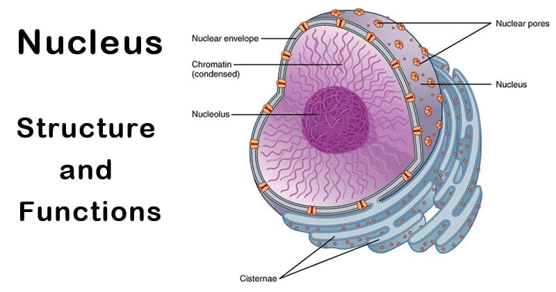
Nucleus: Definition, Structure, Parts, Functions, Diagram 1. Plasma membrane: a selective barrier which encloses a cell (plant and bacteria cells also contain a cell wall ). 2. Cytosol: located inside the plasma membrane, this is a jelly-like fluid that supports organelles and other cellular components. 3. Cytoplasm: the cytosol and all the organelles other than the nucleus. 4.

Source Image: microbenotes.com
Download Image
Anatomy of a Model Cell, Part 2 Diagram | Quizlet 1. Write the name of the cell part in the box next to its description/function. Site of protein synthesis; attached to the outside surface of the endoplasmic reticulum; produces proteins for use outside the cell or for use in the cell membrane. 2. Indicate if each of the following is true of chromosomes or chromatin.

Source Image: quizlet.com
Download Image
In the diagram below, Cell I and II represent typical cells. In both cells, organelle 5 is the site of – brainly.com
Anatomy of a Model Cell, Part 2 Diagram | Quizlet Students practice labeling organelles on a simple model (2D) and a more complex model. The idea is for students to gain an appreciation for how cell diagrams are created. They don’t all look alike, and are often artistically created. Cell organelles tend to follow basic design rules, like the mitochondria will generally look like a peanut
PARTS OF THE CELL. Label the numbered parts. Then complete the table by writing down the differences 2. – Brainly.ph Nucleus: Definition, Structure, Parts, Functions, Diagram [q] The two following parts aren’t numbered (or even shown) in the diagram, but you should know their functions. Lysosomes are involved with intracellular digestion and recycling of worn-out cell parts. Lysosomes are only found in animal cells. Vacuoles are organelles used for temporary storage.